Thank you for visiting our website.
This site is exclusively reserved for health professionals.

Une professionnelle de santé en train de travailler

I am a health professional

Un patient est au téléphone

I want to access the \“patient\” website

Atrophied Sectors

Banner_Clinical_Atrophied Sectors_Posterior mandibular atrophy

Mandibular bone volume may be reduced due to genetic predisposition or local unfavourable factors

Loss of one or more teeth leads to resorption by the alveolar bone which can sometimes extend to the base bone. The size of this resorption varies according to the number of teeth lost, the volume occupied by the roots and the duration of the edentulous space without prosthetic restoration. It is often significant in the molar region where the roots occupy significant space with respect to bone socket volume.


POSTERIOR MANDIBULAR ATROPHY

If this loss of posterior bite is not corrected, long term risks are malfunctioning of the manducation apparatus, which may even have a long-term aesthetic impact on the face due to mandibular rotation and loss of vertical dimension.

In all cases, whether unilateral or bilateral, a posterior edentulous space immediately leads to mastication discomfort with reduced masticatory potential.


Clinical Experiences_Atrophied Sectors_Posterior Mandibular Scan


TWINKON® IMPLANTS

Clinical Experiences_Atrophied Sectors_twinKon® Implants

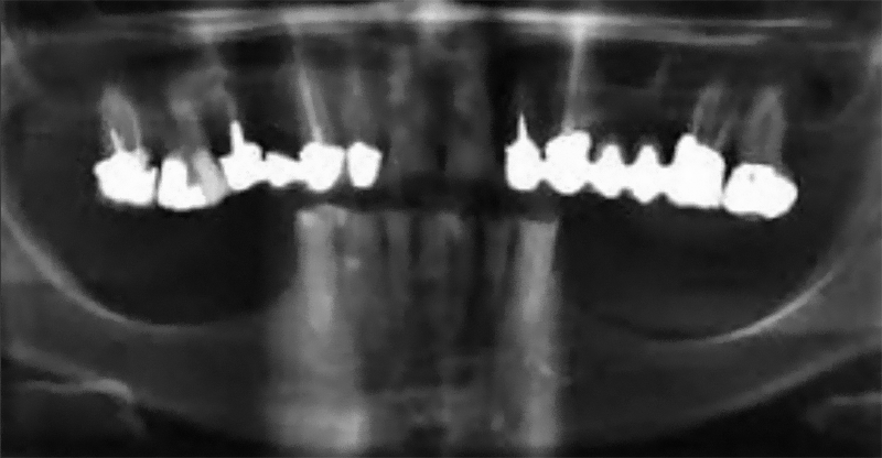
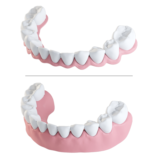
Rehabilitation of mandibular atrophy is a major challenge in oral implantology. Techniques such as vertical bone augmentation grafts or transposition of the dental nerve have been reported but with significant postoperative risks: bone resorption, severe pain or loss of sensitivity(1).
Alternatively, many 8-year studies show that it is possible to obtain satisfactory or better clinical results with comparatively short implants rather than longer implants placed in the augmented bone(2).


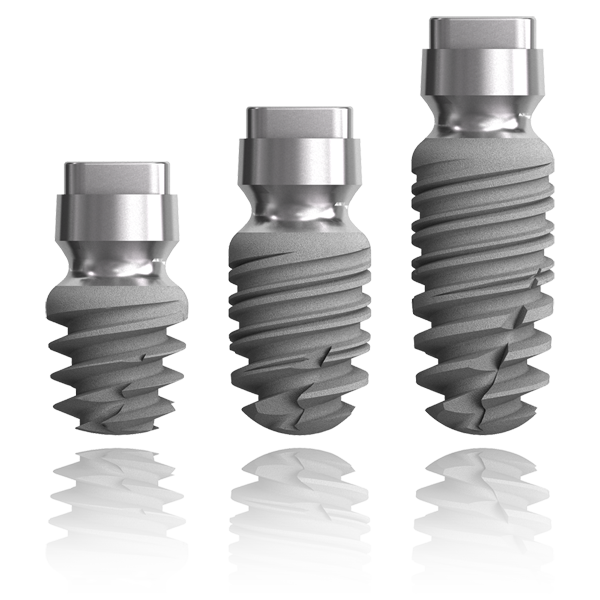
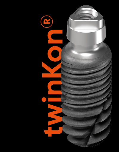
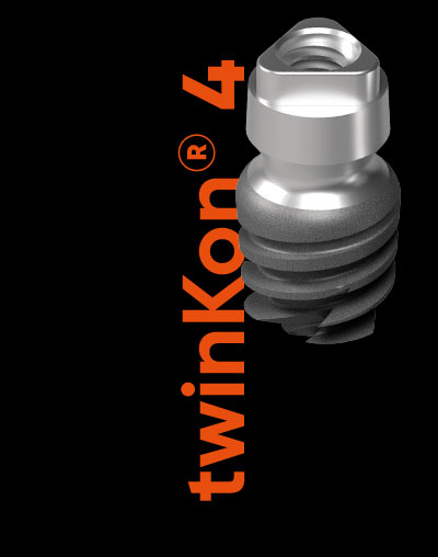
Revised Tissue Level

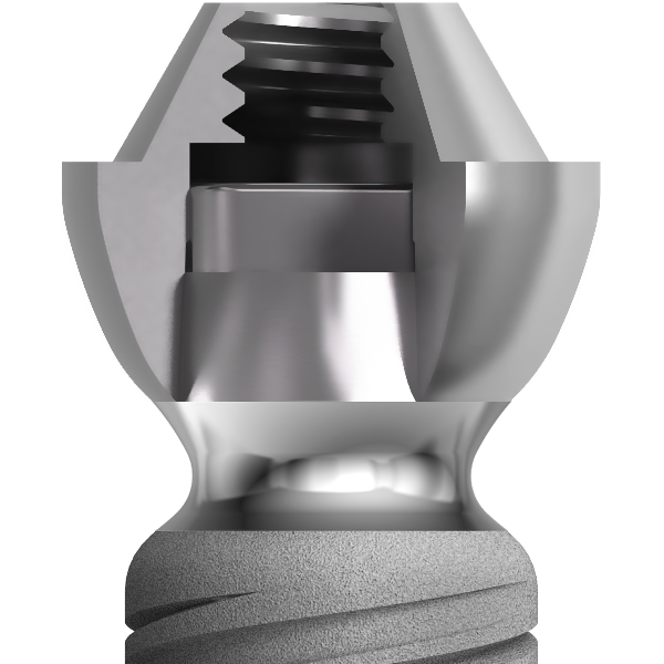
The twinKon® has a rounded shoulder at the limit of the roughened area of the implant. This shoulder is surmounted by a smooth concave collar in contact with the junctional tissue. The aim of the concave collar is first to recreate a mucosal seal which will act as a natural barrier. All subsequent prosthetic manipulation will take place above this limit, therefore reducing the risk of iatrogenic contamination with the benefit of preservation of the residual bone.


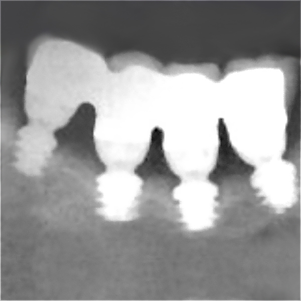
Concave collar and tissue reorganisation

In a study on dogs, Doctor Caroline Bolle demonstrated that after 12 weeks, there was reorganisation of the connective tissue which was rich in fibroblasts and collagen fibres anchored in the bone and converging horizontally on the implant. In the healing space defined by the concavity of the transmucosal collar, circular horizontal organisation of the collagen fibres was observed with a thickness of 500 mm (from the collar to the edge). On a small scale, vertical slices revealed dense circumferential formation of the collagen fibres.

C. Bolle– Early Periimplant Tissue Healing on 1-Piece Implants with a Concave Transmucosal Design: A Histomorphometric Study in dogs (2015)

ULTRA-SHORT TWINKON4® IMPLANT

Clinical Experiences_Atrophied Sectors_twinKon®4 Scan Implants
  • EAO oral communications first prize 2015
  • EAO oral communications first prize 2016

Global D offers a unique, minimally invasive, alternative to vertical bone augmentation. twinKon4 is the L 4 mm x ø 4 mm ultra-short implant of the twinKon® range. This implant can be used as an alternative to L 8.5 mm x ø 4 mm implants in the posterior mandibular area(3).

FIXED TWINKON® PROSTHESIS

Clinical Experiences_Atrophied Sectors_twinKon®_Screw Retained Fixed Prosthesis

Repeat intervention and no cementation

Determining the mode of prosthesis fixing is an important step of the treatment plan. In atrophied regions restored with a fixed prosthesis, it is advised to allow for the possibility of a repeat intervention (maintenance) and avoid using cement (risk of subgingival fusion). The screw retained prosthesis on implants is defined as a screw retained restoration on conical abutments which are themselves fixed to the implants. Thanks to the screw access holes, the screw retained prosthesis is the most comfortable solution in terms of maintenance.

COMPLETE MANDIBULAR FIXED PROSTHESIS

Clinical Experiences_Atrophied Sectors_Complete Mandibular Fixed Prosthesis

Screw retained bridge with false papillae

The prosthesis consists of the crown area and the exposed area of the roots. This restoration can be completed over 4 mandibular implants and 6 maxillary implants. Alveolar loss is moderate with this technique. Note that the prosthesis must provide enough spaces to allow interdental brushes to be used by the patient.


Screw retained bridge with false gum

The prosthesis consists of the missing teeth and a large false gum. This solution is indicated when significant vertical bone loss does not allow treatment with a full implant-supported category 1 or 2 bridge. The false gum enables labial support when this is no longer assured.

ULTRA-SHORT DRILLS WITH DEPTH STOPS

Clinical Experiences_Atrophied Sectors_twinKon®_Ultra-short Drills and Surgery Kit

twinKon 4® Surgery Kit

twinKon®4 ultrashort implants have a specific surgery kit. The kit is supplied with ultra-short drills with fixed depth stops. During boring, the drills are used at a very slow rotation speed to provide optimal control of drill depth and axis. Implants over 4 mm in length are placed using the ULTIMATE kit.

BAR PROSTHESIS

Clinical Experiences_Atrophied Sectors_Bar Prosthesis

Prosthesis for total edentulous patient

A non-moveable prosthesis on implants is planned in cases of moderate to severe resorption of alveolar bone. As well as resolving apparatus mobility problems following resorption of the bone crests, this solution enables regaining stable occlusal relationships, restoring masticatory function and reducing signs of aging by maintaining the facial tissues.

INTEREST OF TISSUE LEVEL IMPLANTOLOGY(4)

Shifting of the abutment-implant interface and tissue stability

  • Simplification of the surgical protocol completed in one surgical step,
  • Shifting of the prosthetic interface over the bone crest,
  • Immediate healing after surgery of soft tissues in contact with the smooth collar of the implant,
  • In-mouth prosthetic accessibility for manipulation.
(1) AR Rokn Comparing 4 mm implant to longer implant pllaced in augmented bones in the atrophic posterior mandibles : one year results of a randomized controlled trial (2018)
(2) Pietro Felice Posterior jaws rehabilitated with partial prostheses supported by 4.0 x 4.0 mm or by longer implants: Three year post-loading results from a multicentre randomised controlled trial (2018)
(3) Pietro Felice Posterior jaws rehabilitated with partial prostheses supported by 4.0 x 4.0 mm or by longer implants: Three-year post-loading results from a multicentre randomised controlled trial (2018)
(4) Information Dentaire n 27 17 juillet 2019 (page 68 74)